


The vertebrae are located in the midline of the body, which is joined to the head by the top and bottom by the pelvic bones and consists of 26 distinct bones. The vertebrae comprise and support the spinal cord and consist of four sections:
تیترهای مهم این مقاله
What are the parts of the spine?
Cervical (7) / Thoracic (12) / Lumbar (5) and 5 Sacral

Spinal anatomy
Intervertebral disc
Between each vertebra there is a disc ,discs are elastic pads that are mounted to strike and facilitate the movement of the vertebrae, consisting of a gelatin core(Nucleous) and an outer wall. Lumbar disc herniation occurs when the gelatinous material of the disc (nucleus) protrudes from the outer wall of the disc, which can be due to age / trauma / weight, etc. As in the lower figure, the protruding disc puts pressure on the spinal cord and nerve fibers, causing pain in the spine and organs and necessitating surgery.
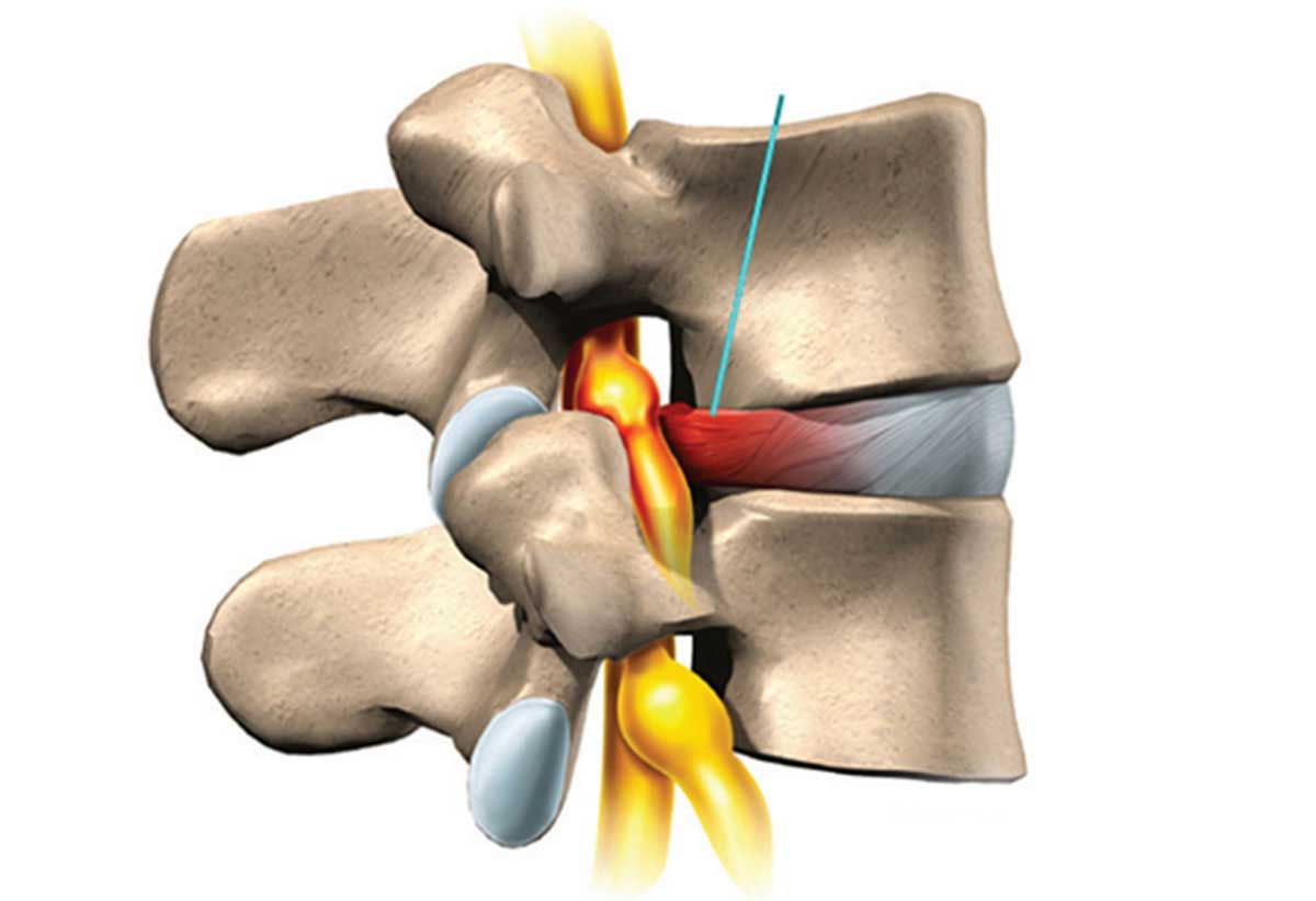
Lumbar Disc Herniation
Disc surgery has two forms
1- Open laminectomy :
The old procedure, which is open surgery, in which the spinal cord tissue is opened and the site of the stenosis is shaved and the nerve is released. The open surgical procedure has complications, the most common of which are: a/ general anesthesia complications b/ complications of surgical manipulation including damage to adjacent tissues and infection c/ postoperative epidural adhesions that can be make nerve pressures and produce pain. d/ Vertebral instability and pressure and osteoarthritis of the intervertebral facet joints.
2- Minimally invasive intradiscal procedure :
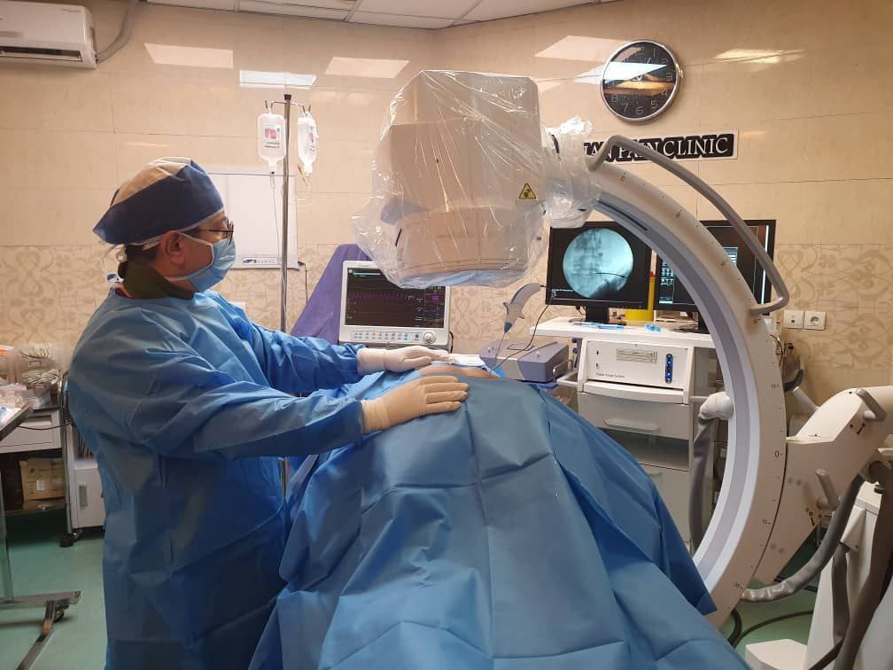
Because of the complications of open surgery mentioned above, closed disc surgery or minimall invasive procedures invented. In a closed surgery, the tissue is not opened and local anesthesia is performed, with special equipment being inserted into the disc protrusion site and the disc herniation removed.
Intradiscal disc decompression procedures are performed by Dr. Mohammad Reza Kazemi, a pain specialist in the following two ways:

Laser operation of closed PLDD disk
A/Percutaneous laser disc decompression (PLDD)
In this procedure, a special needle is inserted into the center of the disc by fluoroscope or CT Scan guidance and a laser fiber is directed through the needle to the center of the disc. The protruding part of the disc (which presses on the nerve) shrinks and the mechanical pressure on the nerve decreases. This is done only on small disc herniation that have a good disk height and the disk core water is not lost.
-
- انجام عمل اندوسکوپی دیسکEndoscopic Discectomy
-
- Endoscopic Discectomy
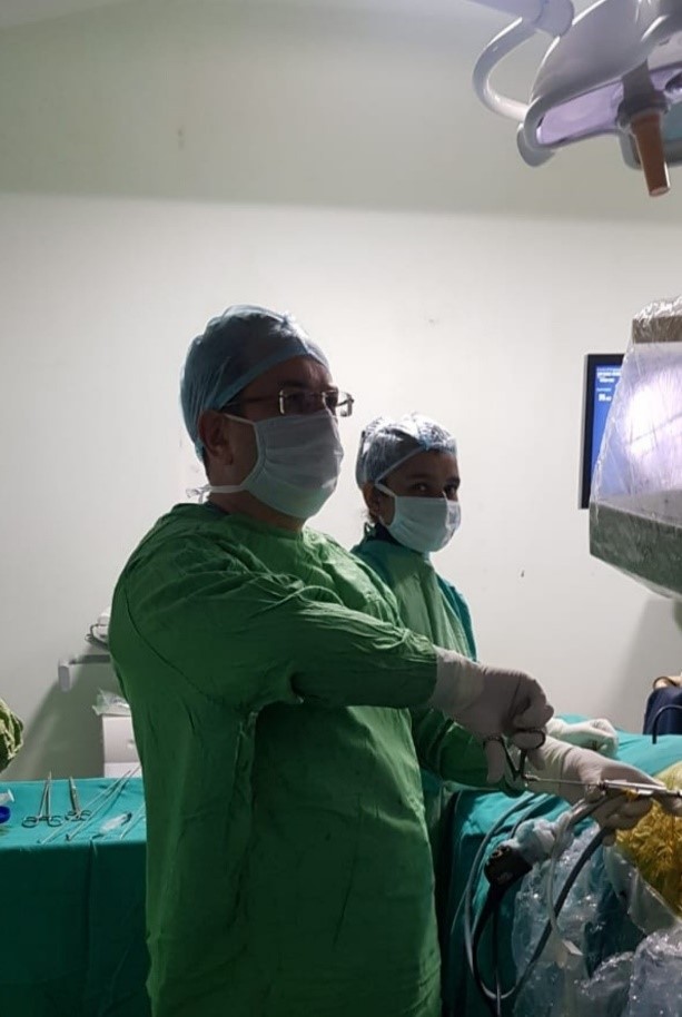
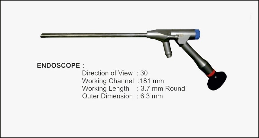
B/Endoscopic Discectomy
An endoscope is a device that allows the vertebrae and tissues around the disc to be directly seen by means of a small incision (within a few millimeters) in the desired area (Lumbar, neck …). One can remove a part of the disc that causes pressure on the nerve (figure above). In this operation, the endoscope with a special camera and light source is inserted into the protruding disc with a small incision of a few millimeters. The disc is also repaired with radiofrequency. It is evident that due to the lack of tissue manipulation the usual open surgical complications are not seen in this technique.
All surgical procedures (interventional) are with local anesthesia and the recovery time is very short.


